
Life Sciences Centre
2350 Health Sciences Mall
Vancouver, British Columbia
Canada V6T 1Z3

Neuronal specification during nervous system development and maturation in Drosophila
Complex nervous system function depends upon the generation of many different subtypes of neurons and the lifelong modulation of their function by intercellular communication. The identity and function of a particular neuronal subtype is a product of the unique repertoire of ‘terminal differentiation’ genes that it expresses (eg. guidance molecules, ion channels, receptors, neurotransmitter biosynthetic enzymes, neuropeptides etc).
These genes are turned on after the neuron is born, but how neurons turn on the right set of genes is still poorly understood. Two regulatory inputs have been shown to play a role in regulating the expression of such ‘terminal differentiation’ genes; 1) the unique code of transcription factors expressed within the neuron, and 2) factors/signals that are secreted from the target cell that the neuron innervates. Our studies in Drosophila have demonstrated that these two inputs may actually functionally intersect to determine the mature gene expression repertoire of neurons.
Our research aims to investigate the mechanisms by which neurons selectively express the genes that define their unique identities and functions, exploring the roles of combinations of transcription factors and extrinsic signals. The genes and mechanisms that govern cell specification are highly conserved from invertebrates to humans. Therefore, we can learn much about the fundamental principles and molecular mechanisms relevant to vertebrate neuronal differentiation using the simpler nervous system and genetic amenability of Drosophila.
Disruption of transcription factors has been linked to congenital neurological disorders, and disruption of intercellular communication and trafficking of target-derived signals has been implicated in neurodegenerative disorders. Our studies will provide a mechanistic understanding of how these factors control gene expression pertinent to neuronal function, advancing our understanding of the aetiology of neurological disorders.
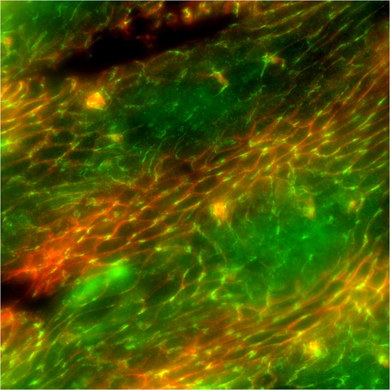
Our research program investigates the roles that glia play in the development and function of the nervious system. Glia are known to fulfill a number of important functions during nervious system development. Glia help guide axon guidance, separate axons bundles in nerves and finally arap and insulate their associated axons and nerves. However many of the molecules and signals that mediate these roles of glia have yet to be determined. This important gives the essential role that glia play in the nervous system of all animals. Any mutation or disease that disrupts glial cell function or development results in disruption of nervous system function and can lead to apralysis or death of the animal.
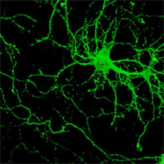
Synapses are the essential point of contact between neurons and their targets for the directional flow of information in the nervous system. The study of how synapses form and how they function is fundamental to our understanding of nervous system connectivity and communication. It is now believed that deficiencies in synaptic function are central to many psychiatric and neurodegenerative diseases such as schizophrenia, Alzheimer’s, Parkinson’s and Huntington’s disease. Thus, it is anticipated that a better understanding of the molecular mechanisms that control these highly specialized structures holds great promise for the development of urgently needed, novel therapies for these diseases.
The principle research objective of my laboratory is to elucidate the cellular and molecular mechanisms underlying the formation, stability, and elimination of CNS synapses. We primarily utilize cultured hippocampal neurons as a model system, and extend these studies to genetically modified mouse models when appropriate. Fundamental questions addressed in the lab include; 1) how does cell-cell contact result in the assembly of pre- and postsynaptic compartments, 2) what are the contributions of pre- and postsynaptic elements to the integrity of the synapse, 3) what are the transsynaptic signals that regulate synaptic plasticity, and 4) is synapse elimination a stereotypical process and, if so, what is the sequence of molecular events underlying synapse disassembly?
Answers to these questions will not only reveal mechanisms underlying developmental and neurodegenerative disorders, but will also provide insight into the molecular signals involved in synaptic strengthening, a process believed essential for learning and memory.
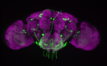
Our brains are composed of billions of neurons, wired together in neural circuits that process information from the environment and produce behaviours. My lab is interested in the organization, function, and development of these circuits. We study this problem in the fruit fly Drosophila melanogaster, an organism with a brain that is much simpler than ours (~100,000 neurons compared to our ~100 billion), but still capable of generating complex behaviours. The fly also offers a powerful array of molecular and genetic tools for identifying, manipulating, and measuring the activity of neural circuits. With a focus on the circuits underlying taste perception and feeding behaviour, we are interested in the following questions:
Our hope is that answering these questions will reveal fundamental principles of neural circuit assembly and function, and important molecules that regulate feeding. Since many of the characteristics of fly circuits are likely to be conserved in mammals, this should give us insight into our own brain, and how it controls what (and how much) we eat.
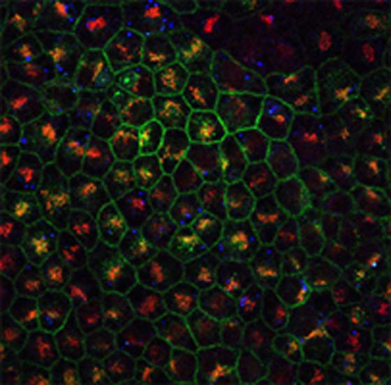
Researching the role of dystroglycan, a protein associated with several forms of muscular dystrophy, in the central nervous system. Molecular mechanisms underlying the dystroglycan-mediated targeting and polarization of proteins in glial cells.
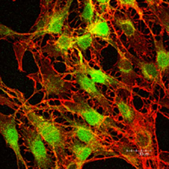
Gap Junctions in Neural Development and Disease
Gap junctions are collections of intercellular membrane channels that join adjacent cells in every organ of the body. They allow a variety of small molecules to pass freely from cell to cell, coupling the cells metabolically and allowing them to coordinate their responses to various signals. The importance of gap junctions has become evident with the identification of congenital diseases resulting from mutations in connexin genes, including X-linked Charcot-Marie-Tooth disease, congenital cataracts, deafness, heart defects and skin diseases. In addition, reduced gap junctional coupling between cells has been detected in several cancers, and increased coupling has implications for epilepsy and stroke. Most of these disease syndromes, to greater or lesser extent, are reproduced in transgenic mice lacking specific connexins.
The objective of my research program is to explore the role of gap junctions in neural development and disease, including consequences of connexin mutations on gap junction structure and function, and to explore the role of these intercellular channels in diagnosis of disease and development of novel therapeutic strategies.
My research in developmental neuroscience is aimed at exploring the function of gap junctional coupling in the developing brain, using pharmacological manipulation as well as genetically modified mice designed to express normal and mutant connexin genes with specific temporal and spatial expression patterns. The role of gap junctions in the etiology and possible therapy of neurological disorders is being examined in animal models and clinical tissues related to stroke, epilepsy and brain cancer. In the area of cell biology and cancer research, we have shown that tumour cells engineered to re-establish gap junctional communication show suppression in growth and tumorigenesis. A major focus of ongoing research is aimed at determining the mechanisms underlying this tumour-suppressive effect, using genomics approaches to identify some of the links between gap junctions and expression of growth control genes. We are also exploring the repertoire of endogenous molecules which pass through gap junction channels, some of which are likely to be involved in the control of cell growth and differentiation. Given the evidence that some tumour therapeutic agents readily pass through gap junctions to enhance tumour cell killing, this research is particularly relevant to the development of novel cancer therapies.
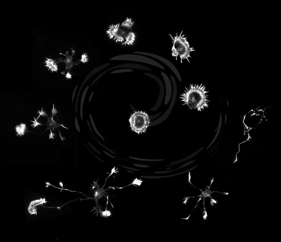
My lab is currently working on three main projects
(1) Intracellular signaling during neurite outgrowth and sprouting. The aim of this research is to identify the intracellular signaling mechanisms important for neuronal outgrowth and to determine their effect on the cytoskeletal network. Recently we have shown that axon consolidation is an active process and that neurite sprouting is suppressed along the length of a neurite. We are currently examining the regulation of this signaling mechanism and identifying approaches to inhibit the signaling and stimulate neurite sprouting. In addition, we are also examining how the cytoskeleton changes in a neuron as it grows in its normal embryonic environment. Using a model insect system of neuronal growth, we analyze the location and activity of key regulators of cytoskeleton function in order to assess how neurons grow and turn in response to embryonic guidance cues. Using sophisticated imaging technologies we will provide some of the first observations of cytoskeletal dynamics of growing neurons in their embryonic environment.
(2) Identification of small molecules that stimulate neurite outgrowth and regeneration. We have recently established a high throughput screen to identify novel compounds that will stimulate neuronal growth and regeneration on inhibitory substrates. Our goal for this project is to test these molecules in animal models as potential ther apeutics to stimulate neuronal regeneration and sprouting in the injured spinal cord. In the future we hope to similarly examine whether these molecules can provide therapeutic benefits to neurodegenerative disease models.
(3) Examination of the role of semaphorins during embryonic development. The aim of this project is to determine the function of semaphorins in the developing nervous system. Semaphorins are the largest class of guidance cue that is expressed in the developing nervous system and many of these molecules have been shown to repel or inhibit neurons as they grow. We are particularly interested in the biochemical regulation of semaphorin function and the receptors and second messenger systems that are stimulated by semaphorins. In addition, we are interested in examining the dynamic distribution of semaphorins, particularly with respect to the distribution of the functional regions of the semaphorin protein. Presently we are working on Semaphorins 1, 2 and 5.
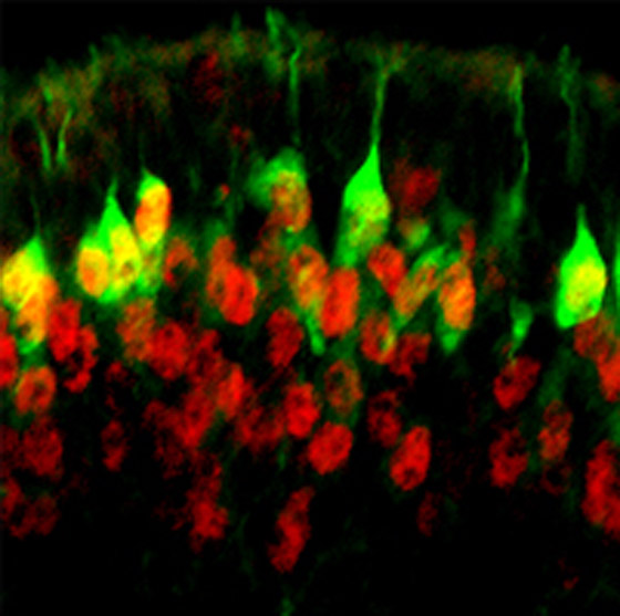
Our lab is interested in the regulation of normal and abnormal nervous system development, and in determining how understanding these mechanisms may be exploited to stimulate regeneration when cells of the nervous system become injured. Many different types of neurons and glia co-exist throughout the brain, however, and their genesis, at different times in different brain regions, creates a confusing milieu in which to study events that occur early in embryogenesis. To overcome this, most of our discovery research has historically utilized a simple, fascinating and uniquely talented part of the nervous system – the olfactory system.
The olfactory neuroepithelium (OE) is one of the most primitive parts of the nervous system. It contains Olfactory Receptor Neurons (ORNs) which are the only recognized projection neurons capable of successfully replacing themselves and re-targeting their axons from the peripheral to central nervous system in a mature animal. In the last 10 years, our research in the olfactory system has revealed novel types of olfactory stem cells, novel mechanisms used by transplanted olfactory-based glia to mediate spinal cord repair, and different ways we may be able to prevent neuronal death in the developing and injured nervous system. All the research we currently perform looks beyond the olfactory system into the brain and is aimed at understanding (1) Neural stem cell regulation; (2) Glia-based mechanisms of stimulating regeneration and brain repair and (3) How the DNA of cells within our brain becomes increasingly more specialized as we stimulate our brain (epigenetic regulation of brain development and function).
The Lab is split into 3 research groups focused on each of these questions, whose interests become more intertwined the deeper we probe into some of the intrinsic mechanisms common to each.
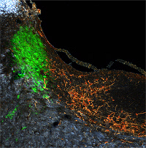
The hypothalamic-pituitary-adrenal (HPA) axis is an important hormonal system in man and rodents, which ultimately controls secretion of glucocorticoids from the adrenal gland: cortisol in humans, and corticosterone in rats. Stress-induced activation of the HPA axis resulting in acute elevations in circulating glucocorticoids levels protect the organism from physiological insult by regulating a variety of physiological processes. For example, they provide adequate substrate for increased metabolic need and help to sustain blood pressure and depress immune function. On the other hand, chronic elevations in glucocorticoids produced by repeated stress exposure have been implicated in the pathogenesis of several forms of systemic, neurodegenerative, and affective disorders.
We have evidence showing that testosterone acts centrally to inhibit stress-induced HPA activity and corticosterone release in the rat. Using a functional neuroanatomical approach assisted by tract-tracing, in-situ histochemical, and early-gene techniques, our goal is to reveal the routes, neurotransmitters, and cellular mechanisms by which testosterone alters circuits in the brain conveying stress-related information. Because gonadal steroid release in both males and females varies as a function of reproductive and social status, this research will lay the groundwork for future studies aimed at understanding the central bases of social- and gender-based differences in stress reactivity.
Life Sciences Centre
2350 Health Sciences Mall
Vancouver, British Columbia
Canada V6T 1Z3